|


|
|||||||||||||||
|
|||||||||||||||

| Investigators: | Christopher Fang-Yen, Mark C. Chu, Seungeun Oh, Ramachandra R. Dasari, and Michael S. Feld |
| Department: | Spectroscopy, Massachusetts Institute of Technology |
We are developing novel interferometric techniques for measuring mechanical and optical signals which accompany electrical activity in nervous tissues. Intrinsic changes include swelling [1], refractive index changes [2, 3], and hemodynamic changes [4]. The potential advantages of intrinsic imaging over electrophysiological methods include the ability to perform long-term noninvasive studies, the ability to record from very small diameter processes (axon/dendrites) for which electrophysiology is impossible, and recording from large numbers of positions simultaneously. In addition, interferometric imaging does not suffer from photobleaching or phototoxicity effects of voltage-sensitive and calcium-sensitive fluorescent dyes.
 |
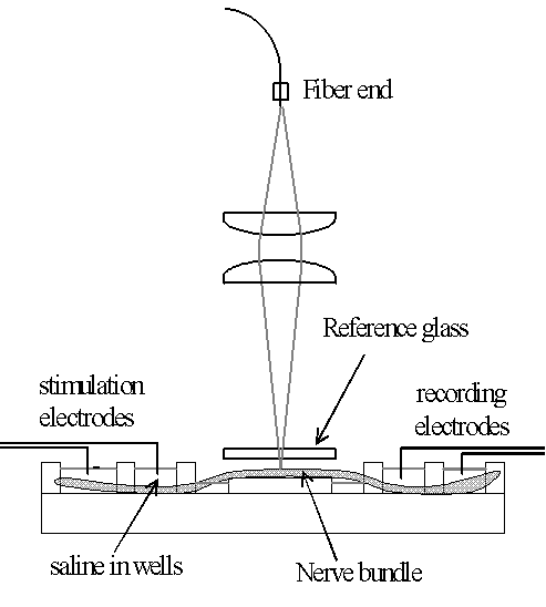 |
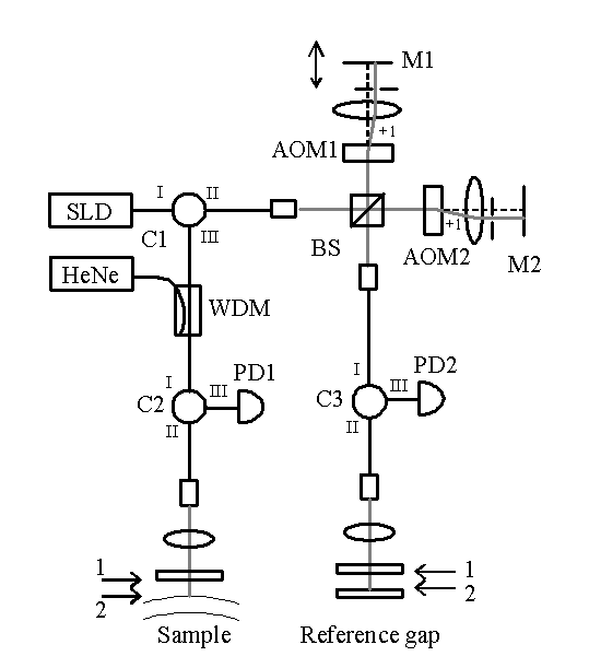
Figure 1a . Interferometer design. SLD: superluminescent diode (center wavelength 1510 nm, 3dB full width 30 nm), AOM1, AOM2: acousto-optic modulators, M1, M2: mirrors; C1-C3: optical circulators; BS: beamsplitter; PD1-PD2: InGaAs photodetectors, HeNe: guide laser, WDM: wavelength division multiplexer; 1, 2: surfaces of sample and reference gap, described in text. Figure 1b . Nerve chamber setup. |
|
We designed a novel dual-beam heterodyne low coherence interferometer [5] to measure nanometer-scale displacements in nerves during the action potential. The interferometer uses a phase-referenced interferometry design, in which the phase of light reflected from a sample is measured relative to a fixed reference reflection [6]. A unequal-arm length Michelson interferometer containing acousto-optic modulators is used to compensate for the path delay between sample and reference reflections. Using this interferometer we performed single-shot, non-contact measurements of the ~5 nm surface displacements during the compound action potential in a lobster nerve. A probe-based version of the interferometer was used to image surface profiles and nerve displacements without a separate reference surface [7].
 |
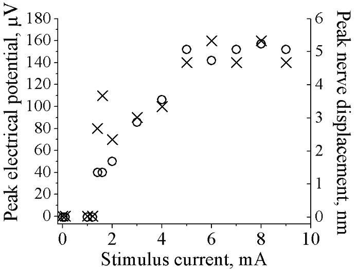 |
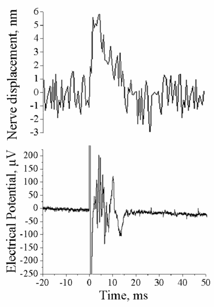
Figure 2a. Interferometrically measured surface displacement and electrical potential of excised lobster nerve. ~5 nm surface displacements observed during action potential. Figure 2b. Peak electrical (crosses) and displacements (circles) for single nerve, with variable stimulus current amplitude. The electrical and optical signals display almost the same threshold and saturation, strongly suggesting the displacements are due to the action potentials. |
|
We have integrated the dual-beam interferometer with an inverted microscope and are investigating related mechanical motions in cultured rat hippocampal neurons and other cells. Fourier domain interferometry and a polarization-division scheme enables the integration of low coherence interferometry with confocal reflectance microscopy. Imaging of cellular motions on the sub-nanometer scale is performed by scanning the sample or interferometer beam.
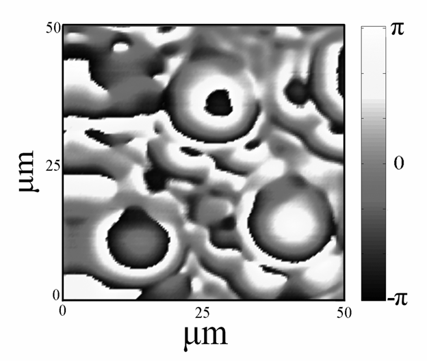 |
Figure 3. Phase image of a portion of a compound eye of a housefly ( Musca domestica ), measured by scanning dual-beam interferometry. Three facets in the fly eye are seen. Curvature of cross section through facet at lower-left corresponds to a radius of curvature 36.8 microns. |
References:
- K. Iwasa, I. Tasaki, R. C. Gibbons, Swelling of nerve fibers associated with action potentials, Science 210 : 338-339, (1980).
- R. A. Stepnoski, A. LaPorta, F. Raccuia-Behling, G. E. Blonder, R. E. Slusher and D. Kleinfeld, Noninvasive Detection of Changes in Membrane Potential in Cultured Neurons by Light Scattering, Proc. Natl. Acad. Sci. 88 (21), 9382 (1991)
- D. Kleinfeld and A. LaPorta, Detection of action potentials in vitro by changes in refractive index, in Light Scattering Imaging of Neural Tissue Function , D. M. Rector and J. S. George eds. (Humana Press, Totowa, New Jersey, 2003)
- A. Grinvald, E. Lieke, R. D. Frostig. C. D. Gilbert, T. B. Wiesel, Functional architecture of cortex revealed by optical imaging of intrinsic signals, Nature 324: 361-364 (1986)
- C. Fang-Yen, M. Chu, H. S. Seung, R. R. Dasari, and M. S. Feld, Non-contact measurement of nerve displacement during action potential with a dual-beam low coherence interferometer, Optics Letters 17, 2028 (2004)
- C. Yang, A. Wax , M. S. Hahn, K. Badizadegan, R. R. Dasari, and M. S. Feld, A phase-referenced interferometer with sub-wavelength and sub-Hertz sensitivity applied to the study of cell membrane dynamics, Opt. Lett. 26 , 1271-1273 (2001)
- C. Fang-Yen, M. Chu, H. S. Seung, R. R. Dasari, and M. S. Feld, Low coherence phase-referenced probe interferometer for surface profiling and displacement measurements in biology, under review (2005)
| [ Home | Overview | Research | People | Facilities | History | Events | Contact | MIT ] | Lab Intranet |