|


|
|||||||||||||||
|
|||||||||||||||

| Investigators: | Jing Kong |
| Department: | Dept of Electrical Engineering & Computer Science, Massachusetts Institute of Technology |
Carbon nanotubes, long, thin cylinders of carbon, are large macromolecules that are unique for their size, shape, and remarkable physical properties. They can be thought of as a sheet of graphite (a hexagonal lattice of carbon) rolled into a cylinder. These intriguing structures have sparked much excitement in recent years and a large amount of research has been dedicated to their understanding.
We are interested in the optical and electrical properties of this fascinating material, as well as how to be able to produce them in a more controlled way. We synthesize our nanotubes with the chemical vapor deposition method. At the Harrison Spectroscopy Lab, we are building a Raman & Photoluminescence setup with tunable excitation to characterize our nanotube materials.
Projects:
(1) Combining Optical Experiments and Transport Measurements
In carbon nanotubes the optical and electronic properties are strongly correlated due to the presence of the van Hove Singularities in the density of electronic states. The energies of the electronic transitions for many different nanotubes lie within the range of visible light, and therefore it is clear that the electronic properties of semiconducting nanotubes will be greatly affected by emission or absorption of photons with specific energies.
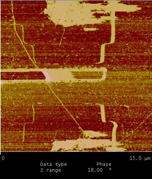
PHOTOCONDUCTIVITY: The excitation of electronic states by a high density of photons can induce carriers in the conduction bands of carbon nanotubes which can contribute to an electrical current. This phenomenon, known as photoconductivity, can be used to probe the electronic band transitions, since the photon absorption is highly increased for photons in with the energy matching that of the electronic transition. Also, a study can be made of electrical contact and structural defect related properties, since the presence of Shottky barriers at the contacts and defects present an intrinsic voltage capable of producing a current with the photo-induced carriers. As shown in Figure 1, even at zero applied bias, photo current are generated when the laser spot is at the nanotube-metal contact region due to the built-in voltage at the Shottky barrier.
 |
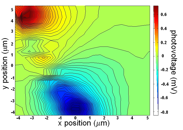 |
Figure 1 (a) AFM image of an isolated nanotube device. (b) Mapping of the photo voltage intensity as a function of the position of the laser spot for the device in Fig.1 (a) at zero bias. The bright red and blue spots correspond to peaks in the photo-current due to the intrinsic voltage from the Shottky barriers at the contacts. |
ELECTRON-PHONON INTERACTION: Phonons are well known to be the greatest contributors for the resistivity of materials. In the case of one-dimensional systems such as carbon nanotubes, the interaction with low energy acoustic phonons is known to be very weak, which results in a quasi-ballistic electron transport at low-biases. In a high-bias condition, the optical phonons, which interact very strongly with electrons, play an important role in the resistivity. The correlation between the density of optical phonons and the transport properties of nanotubes can be explored by combining Raman spectroscopy and transport measurements.
(2) Studying the Electron-Defect interaction
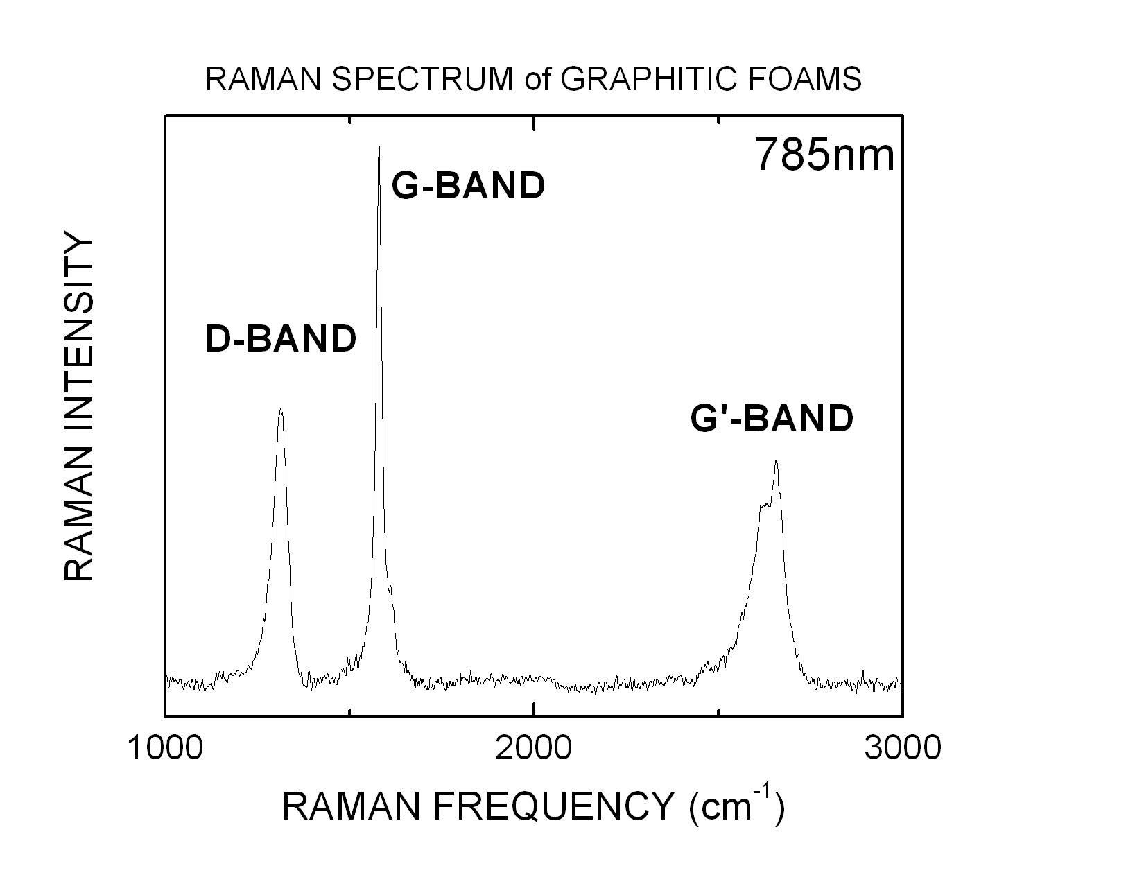
Electrical and thermal conductivity of graphite and carbon nanotubes are greatly affected by the presence of defects. Raman spectroscopy is an important technique for the study of defects in carbon materials due to the presence of defect related peaks, namely the D and G' bands (see Figure 2(a)). The intensity of the defect related features in comparison with non-defect related features is a function of the density of defects and the electron-defect interaction. The dependence of the intensity of the D-band with laser energy can only be attributed to differences in electron-defect interactions. Thus, by probing the relative intensity of the D-band with different laser lines for the same region of graphitic materials (pyrolytic graphite, graphitic foams and carbon nanotubes), the defects can be characterized with respect to their localization (Figure 2(b)).
 |
 |
| Figure 2 (a) Typical Raman spectrum of graphitic materials. (b) Mapping of the D-band to G band intensity ratio vs position in the sample. For x and y positions, the units are in 0.1 m m. The regions with low D-band intensity composed of highly aligned graphite. |
(3) Chirality assignment of carbon nanotubes
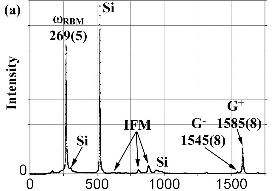
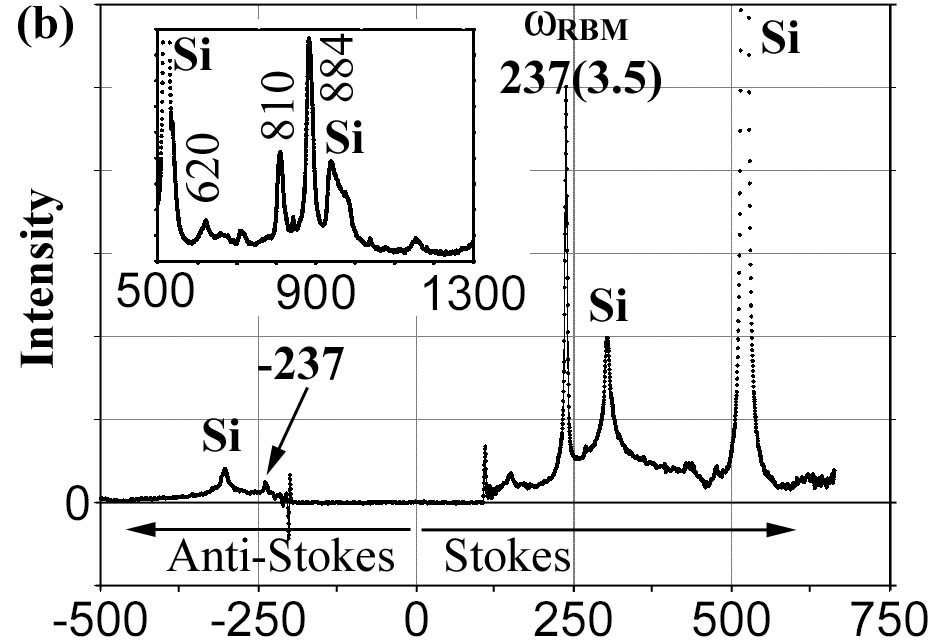
During the past few years Raman spectroscopy has become one of the most commonly used methods to characterize carbon nanotubes. It was demonstrated that under energy resonant conditions it is possible to obtain a Raman signal from a single nanotube and from its Raman spectra this nanotube's chirality can be derived [1]. However, this usually involves an elaborative analysis with uncertainties and only limited number of nanotubes can be probed among many others in a sample. With our tunable Raman setup, we will be able to perform more accurate and straightforward chirality assignment. In addition, we plan to investigate on the relatively un-explored Raman features, such as the intermediate frequency modes (IFM) [2], both to study the scattering mechanisms that give rise to these features but also to use them as a supplemental identification for the chirality assignment. An example is in Figure 3(b) and (c), where the two nanotubes are both under resonant conditions with excitation energy E laser =1.58eV, and both give a radial breathing mode (RBM) frequency of 237cm -1 . However, their IFM features are completely different, indicating that they are two nanotubes of different chiralities.
 |
 |
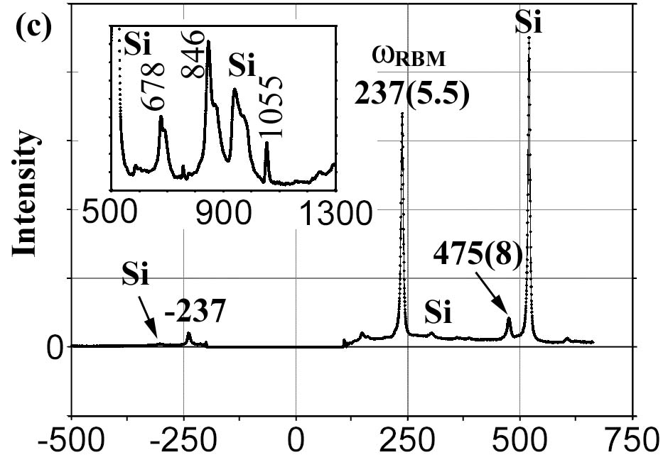 |
| Figure 3 (a) Typical Raman spectra of a suspended single-walled nanotube. The RBM frequency for the nanotubes in (b) and (c) are the same. However, the IFM frequencies and intensities are different, shown in the insets, indicating that the two nanotubes have different chiralities. |
References:
- Jorio, A., et. al. , Structural (n, m) determination of isolated single-wall carbon nanotubes by resonant Raman scattering. Phys. Rev. Lett., 86 , 1118 (2001)
- Son, H., et. al., Environmental effects on the Raman Spectra of Individual Single Wall Carbon nanotubes: Suspended and Grown on Polysilicon. Appl. Phys. Lett. , 85 , 4744 (2004)
| [ Home | Overview | Research | People | Facilities | History | Events | Contact | MIT ] | Lab Intranet |