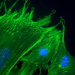
Stained with fluorescein, actin cytoskeletons of human endothelial cells glow green in this immunofluorescent micrograph. DNA stained with DAPI shows blue.
The actin filaments have linked themselves into a highly triangular structure resembling a geodesic dome in these human endothelial cells. Actin takes this form as cells, human endothelial in this case, grip a substrate and spread, forming discrete attachment points on the surface. The geodesic framework functions as a tensegrity structure, giving the cell its architectural integrity. DAPI is a fluorescent stain widely used to visualize DNA.
Image by Sui Huang and Donald E. Ingber, Harvard Medical School.
Source: Donald E. Ingber, "The Architecture of Life," Scientific American, Jan. 1998
In cell biology images often function as primary data and reveal previously unseen events and architectural features. Many examples are presented and discussed at the conference and workshops.