Quantitative analysis and compositional mapping
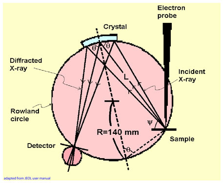
The Wavelength Dispersive Spectrometers (WDS) are used to obtain concentration of all elements present in a spot, and X-ray maps showing distribution of elements in a flat cross-section of the sample. The electron beam diameter is 1 micrometer. Therefore, the spot analyzed is a few cubic micrometers in volume, and the maximum resolution of the X-ray maps is 1 micrometer. WDS takes advantage of Bragg's Law of diffraction, which ensures that only the desired X-ray wavelength reaches the detector. The sample, the diffracting crystal and the detector are positioned on a focusing (Rowland) circle that has a fixed radius by design. The angle of diffraction may be changed to detect other X-ray wavelengths by tilting and moving the diffracting crystal toward or away from the sample. The detector moves simultaneously with the crystal such that the radius of the focusing circle remains constant although the circle itself changes position.
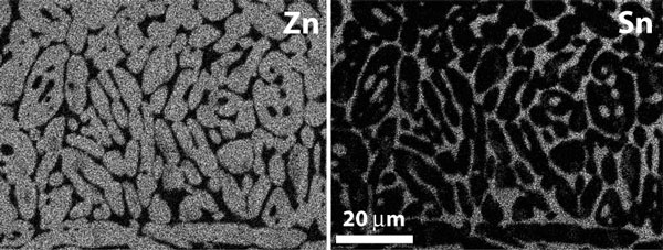
X-ray maps are useful in sets. The number of maps that can be acquired simultaneously depends on the number of WD spectrometers available. There are two methods of acquiring X-ray maps. The first involves a stationary sample as the beam rasters over the desired area. This method is suitable for small area imaging as shown in Zn and Sn images of a Zn-Sn composite below:
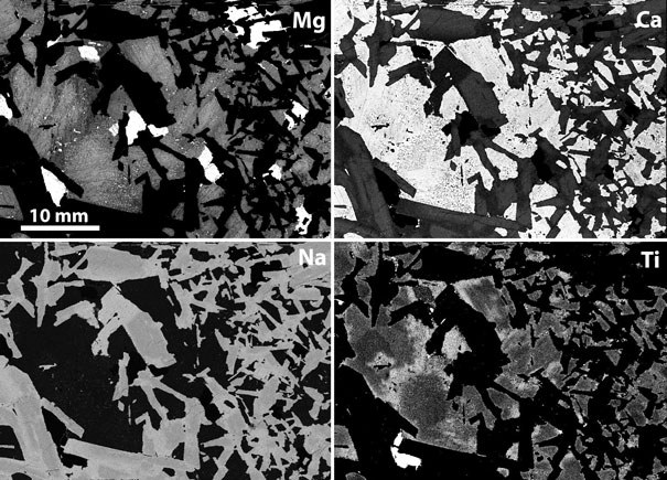
For large areas, however, beam rastering results in defocusing in the peripheral parts of the image. To alleviate defocusing problems, the image is acquired by keeping the beam stationary while the sample stage is moved to expose the surface to be imaged. Four simultaneously acquired maps of a large surface of a rock using this technique are shown below: