Theory
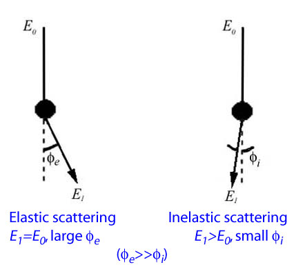
Electron microprobe analysis depends on an electron beam that generates X-rays upon interaction with the specimen. The electron beam is generated by thermionic emission from a tungsten filament or a sharpened lanthanum hexaboride (LaB6) single crystal, and accelerated in a potential difference of 5 to 30 kV. Beam electrons with an initial energy of E0 scatter elastically or inelastically upon encountering a specimen atom and loses a part or all of its energy. High-angle elastic scattering with little energy loss contributes to the back-scattered electron signal used in compositional imaging, whereas low-angle inelastic scattering with significant energy loss results in X-ray emission through inner-shell ionization and subsequent relaxation of the specimen atom. The electron beam must have sufficient energy to cause inner-shell ionization in the specimen atoms in order to generate X-rays.

The intensity of the X-rays are then measured with the Wavelength Dispersive Spectrometers (WDS). Data reduction involves converting measured X-ray intensities to elemental concentrations. This is usually handled by the computer software.


