

Introduction
The Electron Microprobe, also known as the Electron Probe X-ray Microanalyzer (EMPA), is a powerful and well established analytical tool that provides non-destructive, in-situ and complete quantitative chemical analysis of a flat solid surface with a spatial resolution of ~2 micron through X-ray emission spectrometry. It also provides high-resolution scanning electron and elemental X-ray images (concentration maps) showing spatial distribution of elements. A variety of earth, extraterrestrial and artificial materials including minerals, glasses, ceramics, metals and superconductors can be analyzed. Click on the image below for details:


Our JEOL JXA Superprobes are equipped with:
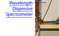
- Five wavelength dispersive spectrometers (WDS) working simultaneously to analyze elements with atomic number >4 (B to U) and minimum detection limits of ~10 ppm under favorable conditions.
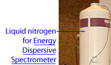
- Energy dispersive spectrometer for qualitative and quantitative analysis of elements with atomic number >8 (F to U).
- Back-scattered electron detector for high resolution compositional and topographic imaging.
- Secondary electron detector for high resolution topographic imaging.
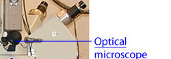
- Light spectrometer for hyperspectral cathodoluminescence imaging and analysis.
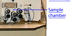
- An orthogonal translational stage that can hold up to nine 1-inch diameter sample mounts or a large sample (up to 80 mm x 80 mm) of irregular shape.
